Friederike Hübner
During my biology studies, especially in the early years, I spent a great deal of time on plant anatomy. Microscopes open up a world of their own, and the cellular structure of a plant tells us much about its adaptations and the environmental conditions at its habitat. I therefore began to examine the genera Ceropegia and Cynanchum in more detail, particularly the stem anatomy of mature shoots.
Here I would like to present the anatomy of Ceropegia linearis subsp. woodii, which in my view is an exceptionally attractive Ceropegia. Its appearance is probably one reason why it has become so widely distributed as a hanging houseplant.
Under good growing conditions Ceropegia linearis subsp. woodii develops quite long internodes, the sections of stem between successive pairs of leaves. The shoot is also comparatively thin. Anyone who has tried to tear such a slender shoot apart will probably have found it more difficult than expected. The reason lies in the cellular construction of the stem.
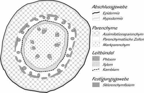
Figure 1 gives a semi-schematic overview of a stem cross-section.
Figure 2 shows an enlarged detail drawn at cellular level.
The outermost layer is the epidermis (“outer skin”), followed by a second complete cell layer, the hypodermis (“lower skin”). Neither layer contains chloroplasts, the green organelles responsible for photosynthesis, but both contain red inclusions that may be lipid droplets.
This is followed by a broad layer of large, chloroplast-rich cells with large intercellular spaces filled with air. Depending on light exposure, some red central vacuoles may also occur. These are large, often coloured spaces inside mature cells, filled with water and waste products; the living cytoplasm itself is reduced to a thin layer along the cell wall.
The vascular system forms a closed ring. On the outside, the phloem – living tissue transporting sugars and other building materials – is protected against excessive shrinkage by very thick-walled cells in cross-section that are extremely elongated and needle-like in longitudinal section: sclerenchyma fibres. These fibres are also responsible for the remarkable strength of the long shoots. The air-free xylem conducts water and transports micro- and macronutrients. Its walls are also relatively thick because transport is maintained almost entirely by tension. This prevents the conduits from collapsing even under severe water stress and helps maintain continuity of the water column. Between phloem and xylem lies the cambium, a layer that produces new rows of cells both outward and inward. This allows the plant to form thicker shoots that can continue to increase in diameter over time.
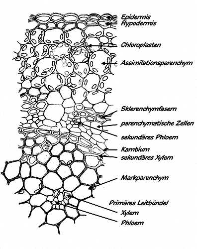
At the centre of the stem lies the pith parenchyma, consisting of thin-walled cells with intercellular spaces. It still contains a few isolated vascular bundles formed during very early growth. Once growth is complete, the plant stores starch grains (leucoplasts) in this pith parenchyma as reserves for “bad times”. I once sectioned a shoot that had not grown for a long period and so many leucoplasts fell out that the cellular structures became difficult to see. This also explains why Ceropegia cuttings taken from old shoots can root relatively quickly: they simply contain ample reserves.